Faculty
Francesco Barca
Country: Italy
Affiliation: SOC Oculistica Piero Palagi Hospital
Webinar


March 12, 2024 - 6.30 PM CET
WATCHInsights into cutting-edge surgical techniques and technologies through reLive surgeries
Videos
 WATCH
WATCH
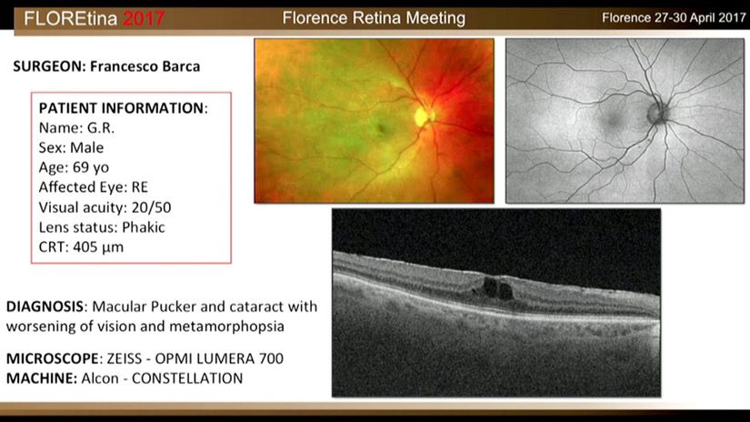
Macular Pucker And Cataract With Worsening Of Vision And Metamorphopsia
Case information:
Male
/
69 years old
- ZEISS - OPMI LUMERA 700
- ALCON - CONSTELLATION
 WATCH
WATCH
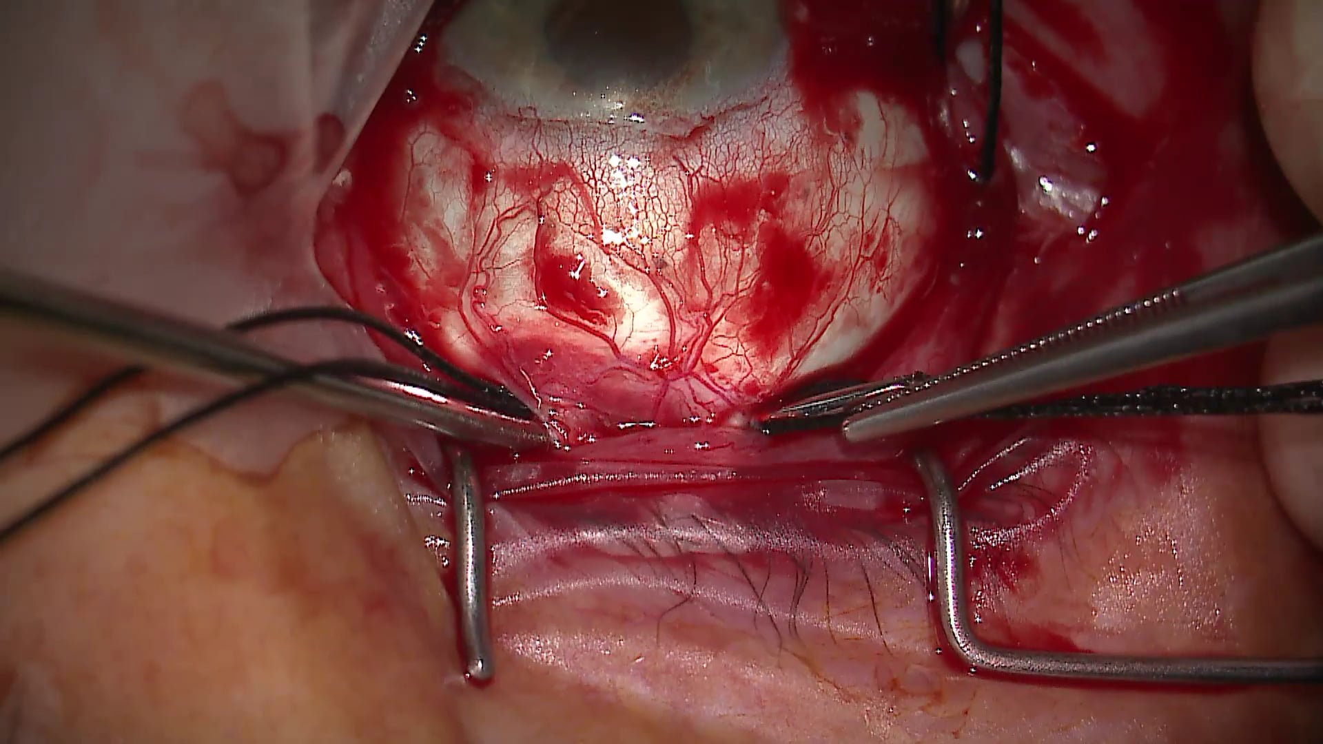
Retinal Detachment
Case information:
Male
/
63 years old
- ZEISS - OPMI LUMERA 700
- ALCON - CONSTELLATION
 WATCH
WATCH
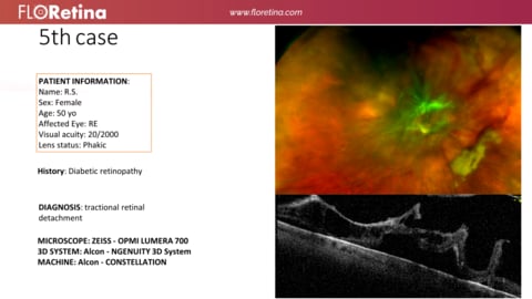
DIABETIC RETINOPATHY
Case information:
Female
/
50 years old
- ZEISS - OPMI LUMERA 700
- ALCON - NGENUITY 3D System
- ALCON - CONSTELLATION
 WATCH
WATCH
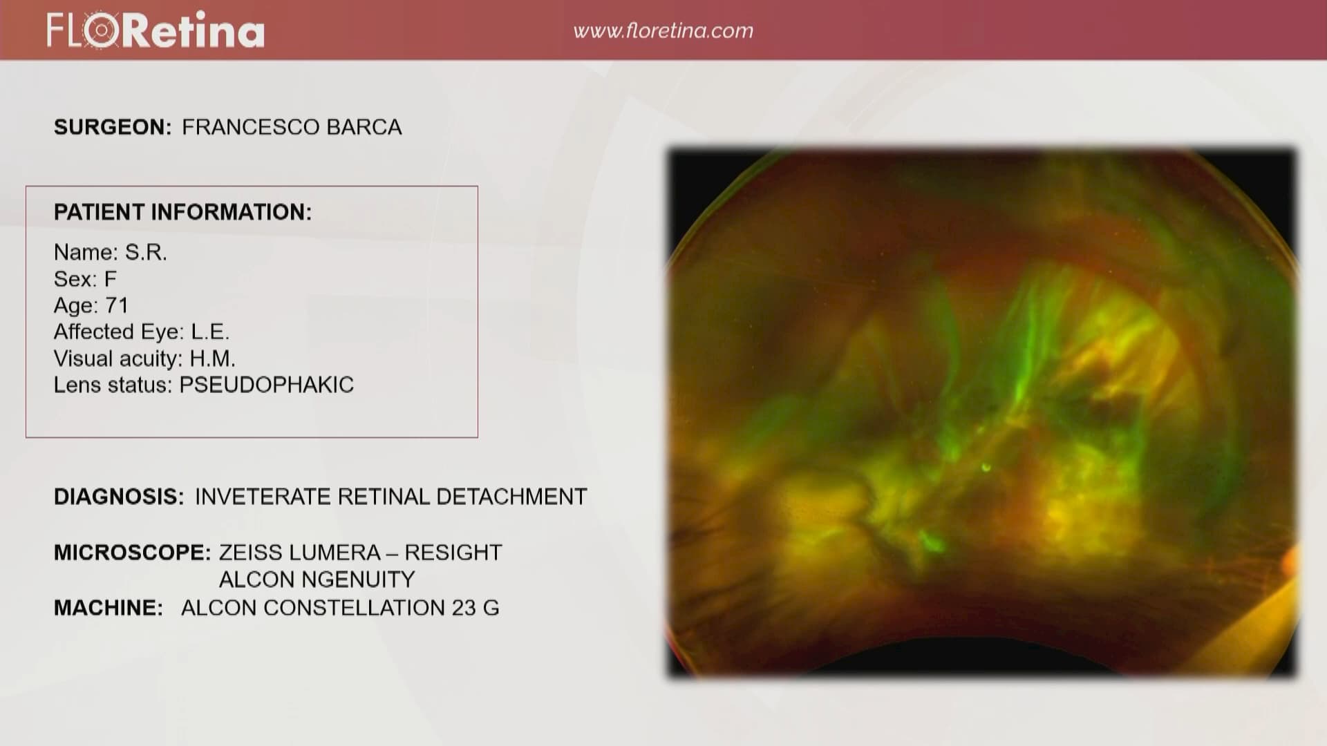
Inveterate Retinal Detachment
Case information:
Male
/
71 years old
- ZEISS - OPMI LUMERA 700
- ALCON - NGENUITY 3D System
- ALCON - CONSTELLATION
 WATCH
WATCH
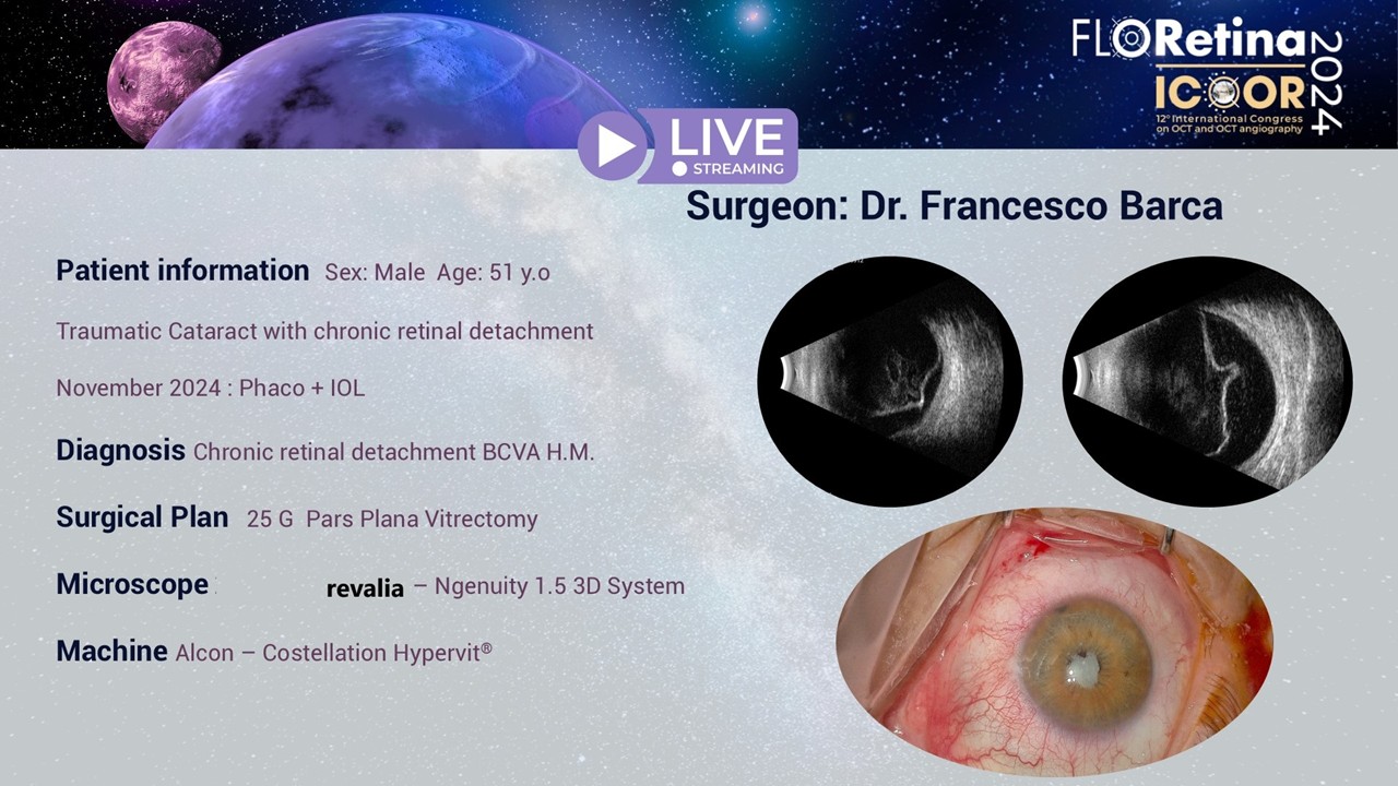
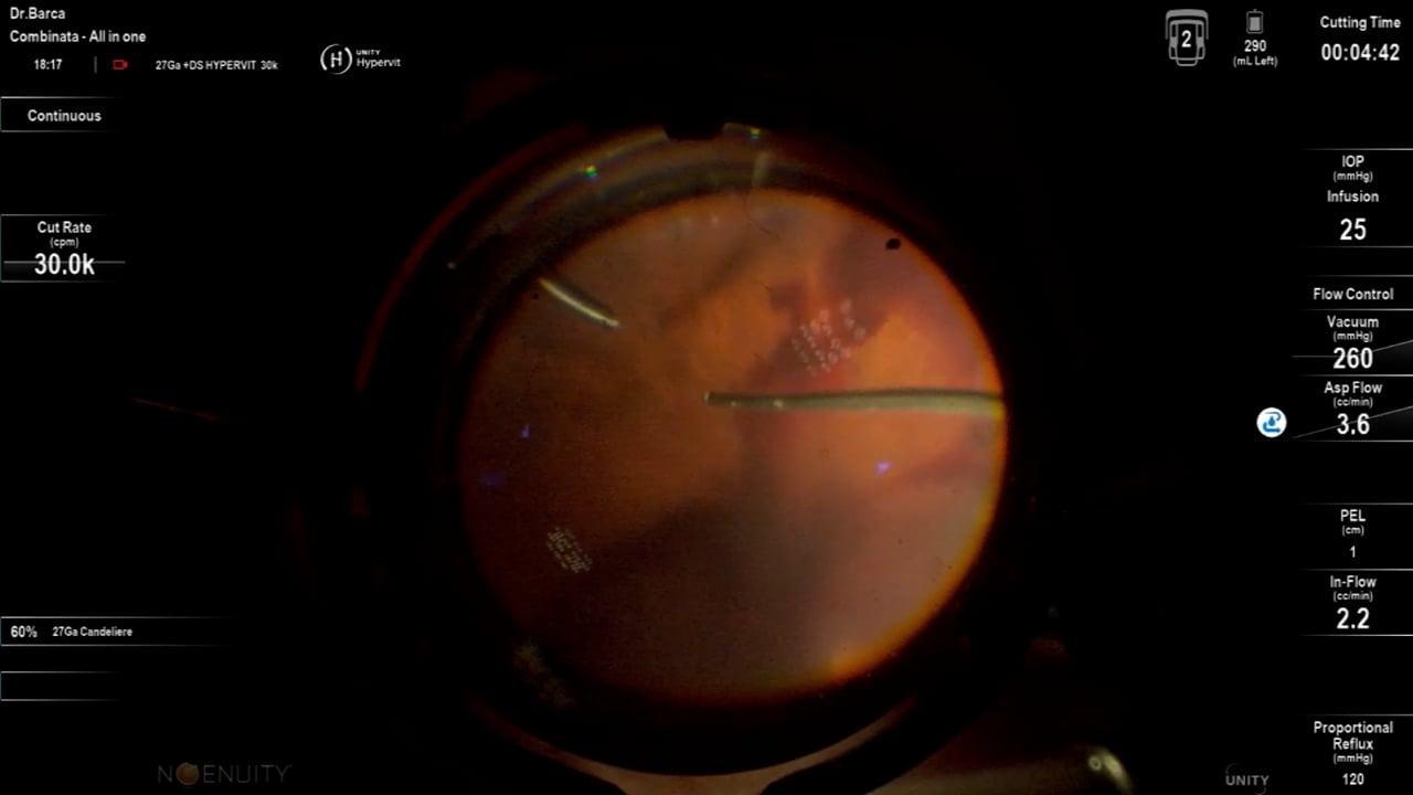
Chronic retinal detachment
- ALCON - REVALIA
- ALCON - NGENUITY 3D System
- ALCON - CONSTELLATION
- ALCON - Hypervit
 WATCH
WATCH
Vitreous hemorrage, secondary to proliferative Diabetic Retinopathy
- ALCON - NGENUITY 3D System
- ALCON - Unity VCS
Image Bank
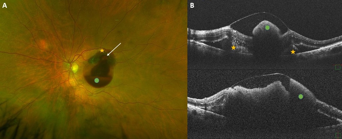
Imaging of a retinal arterial macroaneurysm
Francesco Barca Lorenzo De Angelis Bianca Pacini Mario Galasso Dario Giorgio(A) Ultra-widefield fundus imaging shows a case of a 74-year-old woman with a ruptured retinal arterial macroaneurysm (white arrow) in the left eye. Preretinal (green dot) and subretinal hemorrhages (yellow asterisks) are visible at the posterior pole. (B) Optical coherence tomography examination obtained in the acute phase shows large amount of sub-inner limiting membrane hemorrhage (greendots) with outer-layer shadowing, subretinal hyperreflective material (yellow astetisks) and a pocket of subretinal fluid.
View image
Multimodal imaging of Perifoveal Exudative Vascular Anomalous Complex (PEVAC)
Francesco Barca Lorenzo De Angelis Dario Giorgio Benedetta Pieri Veronica DonatiMultimodal imaging showing PEVAC in the LE of a 73-year-old woman. The patient did not exhibit any clinical signs of diabetic retinopathy, hypertensive retinopathy, retinal vascular occlusion, or myopic degenerative retinal disease. (A) Ultra-wide field fundus imaging showing perifoveal isolated aneurismal lesions ( magnified imaging in the bottom) barely visible on color photography. Drusenoid lesions are detectable in the mid-periphery. (B,C) Fluorescein angiography (FA) reveals well-defined hyperfluorescent lesions well detectable in the (B) early phase with leakage in the (C) late frame. (D) These lesions appear as hyporeflective spots on IR imaging. (E) An increase of the autofluorescence signal is detectable on B-FAF at the macula. (F) Structural OCT examination passing through the lesions showing the presence of an isolated, well-defined perifoveal aneurysmal lesions with a hyperreflective wall (red and yellow arrowhead) and surrounding intraretinal cystic spaces. (G) OCT-angiography confirming the presence of perifoveal capillary abnormalities (green line) in the superficial (red arrow ) and deep capillary plexus (yellow arrow) and a mild rarefaction of retinal capillaries in the perilesional area in both the plexi. No abnormalities are detectable in the avascular slab. More specifically, corresponding B-scans with flow overlay clearly disclose an isolated large dilation (red and yellow arrows) corresponding to PEVAC with detectable flow inside the lesions
View image