Faculty
Tomaso Caporossi
Country: Italy
Affiliation:
Bio:
Medico specialista presso azienda Ospedaliero-Universitaria Careggi Firenze
Webinar


24 May 2022 - 6.30 PM
WATCHThe evolution in vitrectomy probe technologies: easier management of complex surgeries
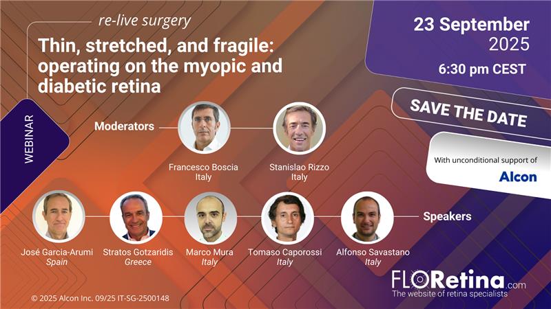
September 23, 2025, 6.30 PM CEST
WATCHThin, stretched, and fragile: operating on the myopic and diabetic retina
Videos
 WATCH
WATCH
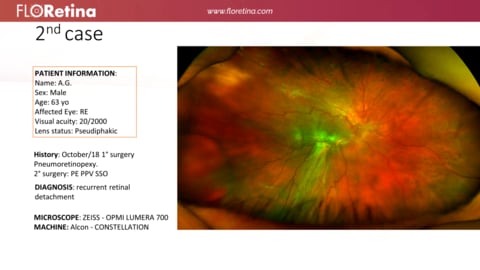
Retinal Detachment
Case information:
Male
/
63 years old
- ZEISS - OPMI LUMERA 700
- ALCON - CONSTELLATION
 WATCH
WATCH
Macular Pucker
Case information:
Male
/
82 years old
- ZEISS - OPMI LUMERA 700
- ALCON - NGENUITY 3D System
- - Resight
- - Oculus Disposable LenZ
- HOYA - Qube Pro 25G
 WATCH
WATCH
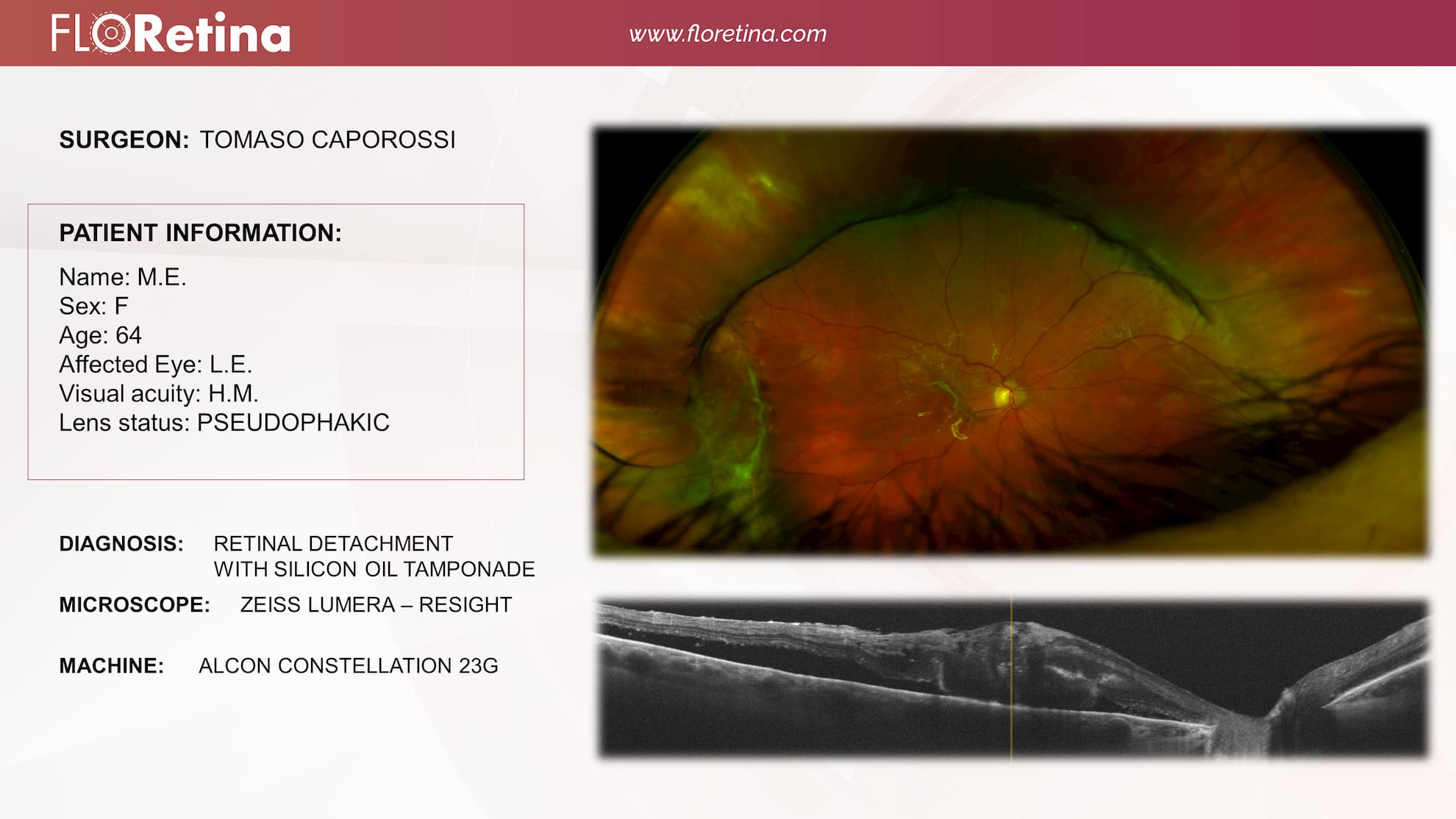
Retinal Detachment With Silicon Oil Tamponade
Case information:
Female
/
64 years old
- ZEISS - OPMI LUMERA 700
- ALCON - CONSTELLATION
 WATCH
WATCH
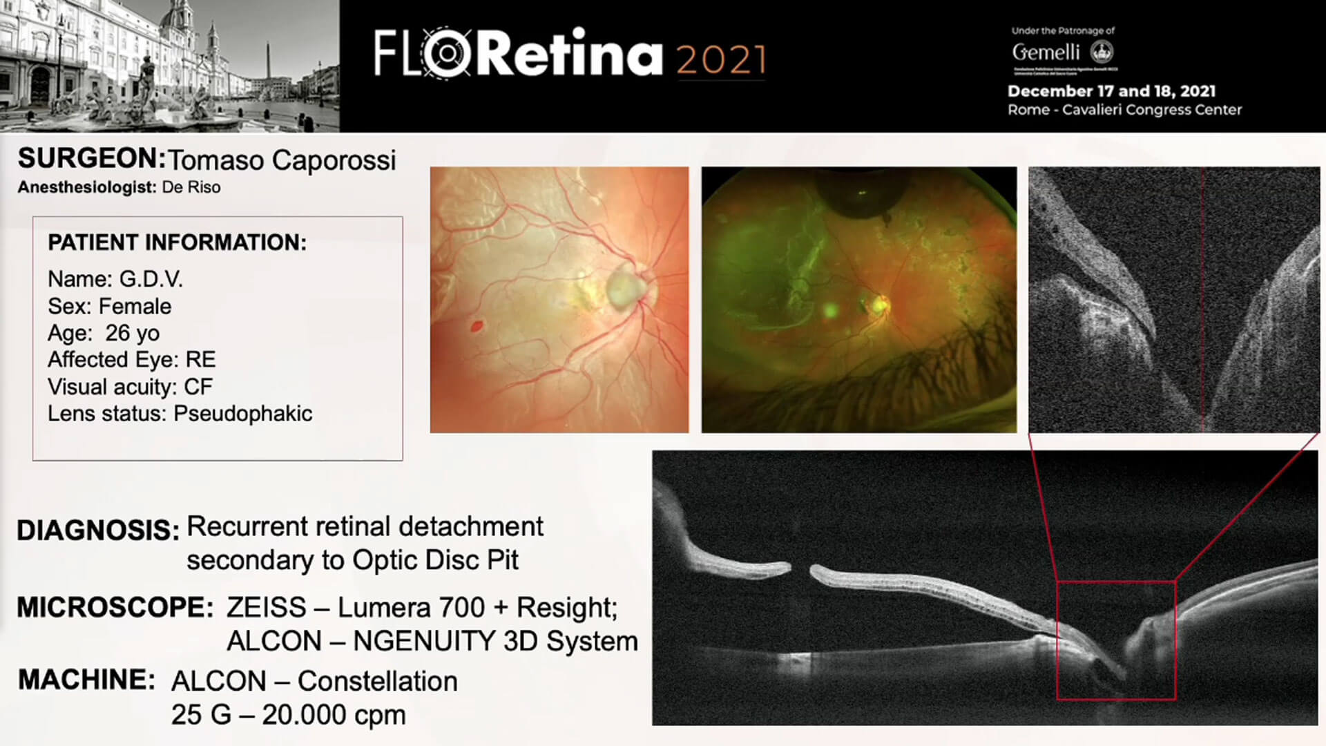
Recurrent retinal detachment secondary to Optic Disc Pit
Case information:
Female
/
26 years old
- ALCON - CONSTELLATION
- ZEISS - OPMI LUMERA 700
- ALCON - NGENUITY 3D System
 WATCH
WATCH
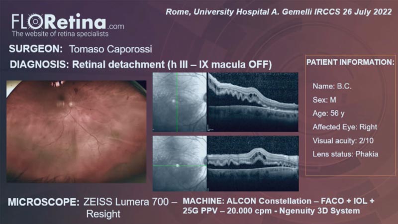
RETINAL DETACHMENT (h III – macula OFF) (Rome, 26/07/2022)
Case information:
Male
/
56 years old
- ALCON - NGENUITY 3D System
- ALCON - CONSTELLATION
- ZEISS - OPMI LUMERA 700
- ALCON - 25G 20.000 cpm
 WATCH
WATCH
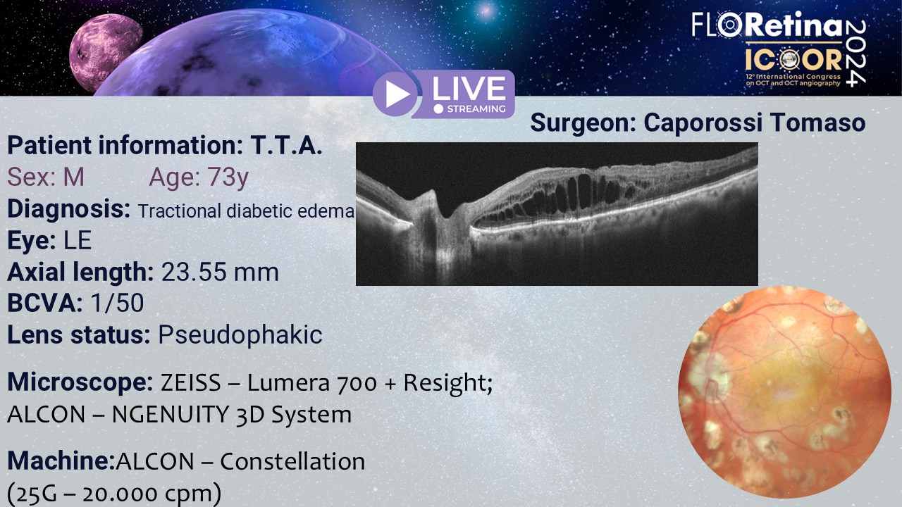
Tractional diabetic edema
- ZEISS - OPMI LUMERA 700
- - Resight
- ALCON - NGENUITY 3D System
- ALCON - CONSTELLATION
 WATCH
WATCH
Recurrent retinal detachment under silicone oil
- ALCON - Unity VCS
- ALCON - NGENUITY 3D System
- ZEISS - OPMI LUMERA 700
- - Resight
Image Bank
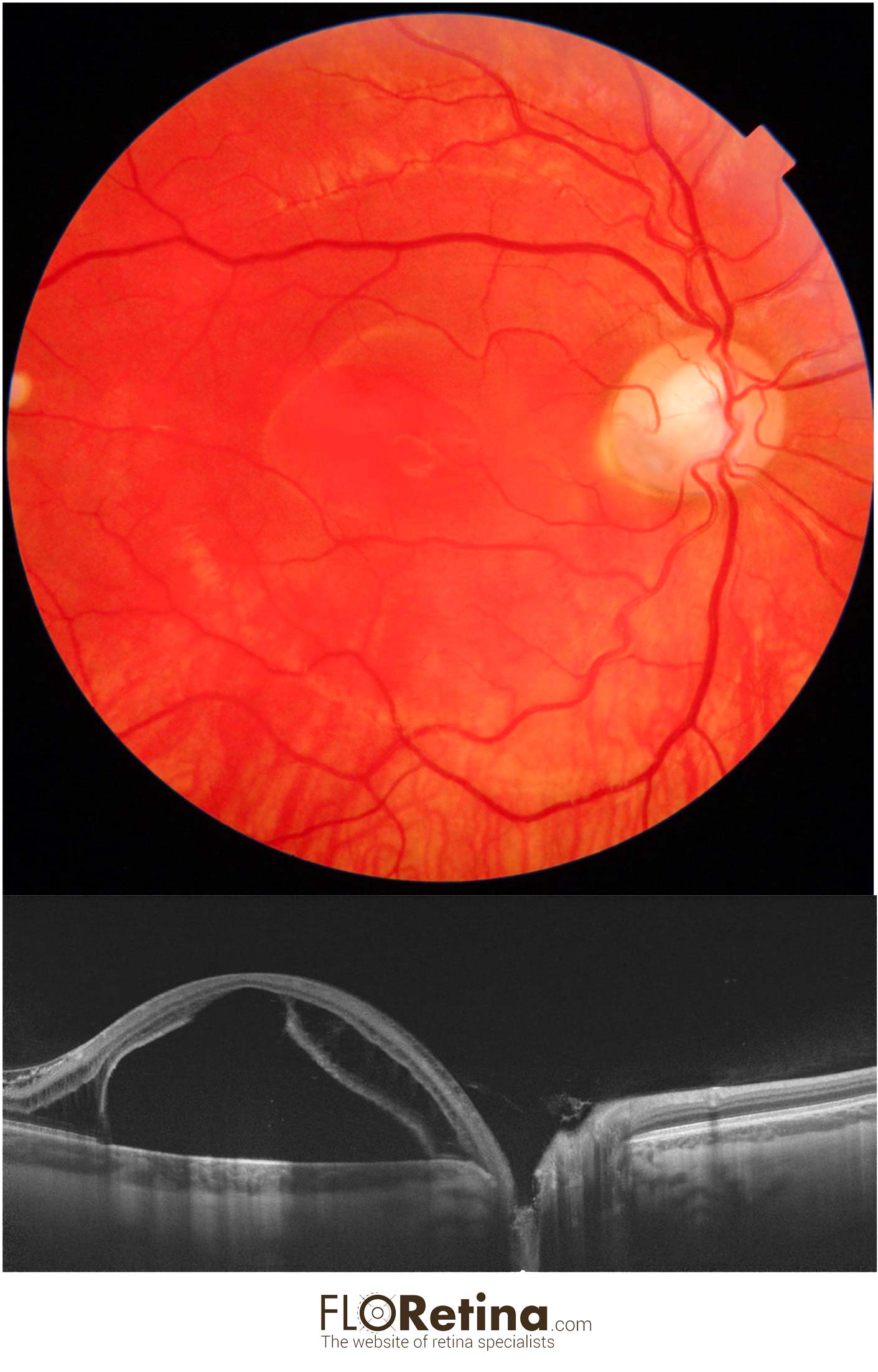
OPTIC DISK PIT
Daniela Bacherini Tomaso Caporossi Alfonso Savastano25 y/o male with visual reduction showing macular schisi with detachment due to optic coloboma.
A. Color fundus photograph
B. Structural OCT showing the retina splitted at both the inner and outer retinal layers with a macular detachment
DEVICE: Fundus photograph (Topcon), Structural OCT (Topcon)